
Dendritic cells (DCs) play an important role in presenting antigens to lymphoid T cells and initiating specific immune responses. The fluorescence properties of Quantum Dots (QDs) make them ideal for two-photon microscopy imaging. Michael D. Cahalan, a member of the University of California, Irvine, used laser confocal microscopy, two-photon microscopy, and electron microscopy to observe the uptake of quantum dots by dendritic cells. It was first discovered that quantum dots regulate the immune function of living organisms.
Debasish Sen et al found that dendritic cells actively take up quantum dots in a manner that relies on the actin skeleton. Initially, the quantum dots entered small vesicles close to the cell membrane; after 10 minutes, vesicles of different sizes, different movement patterns and different fluorescence luminosity were widely distributed throughout the cytoplasm; finally, they were isolated in lysosomes (Fig. 1). Next, in in vivo experiments with transgenic mice, the researchers used a specific antigen protein (ovalbumin Ovalbumin) coupled with quantum dots, and found that quantum dots can act as a high-efficiency antigen-presenting system based on nanoparticles, triggering T cells. activation. Ingestion of quantum dot-coupled antigen-derived dendritic cells induces stable clustering of T cells and up-regulates the expression level of CD69; compared with free antigen, quantum dot-coupled antigen promotes T cell proliferation and interferon production more efficiently (Fig. 2) ). In conclusion, this study is the first to clarify that quantum dots can be used as a nanoparticle-based antigen presenting system to efficiently activate T cells and activate immune response, which is a new application beyond bio-optical imaging.
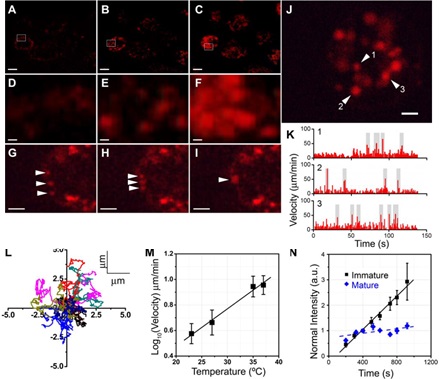
Figure 1. Kinetic analysis of dendritic cells on quantum dot uptake. (AF) Two-photon imaging of quantum dot uptake by dendritic cells at different incubation times, DF is image amplification for AC, respectively; (GI) fusion of vesicles containing quantum dots in dendritic cells; (J) entry into dendrites The quantum dot of the cell; (K) the rate of movement of the vesicle containing the quantum dot in the J-graph; (L) the trajectory of the vesicles of 10 different fractions (tracking time > 140 sec); (M) the fraction of the vesicles The transport is temperature dependent; (N) as the dendritic cells mature, the rate of uptake of quantum dots decreases.

Figure 2 Quantum dot-coupled ovalbumin (QD-ova) activates OT-II T cells in vivo via dendritic cells. (A)-(C) OT-II T cells were immunized with ovalbumin (A), quantum dot-coupled ovalbumin (B), and CFA (C), respectively, and CD69 expression was up-regulated; (D)-(I) Detection of T cell proliferation by CFSE staining: Quantum dot-coupled ovalbumin is more effective in promoting T cell proliferation than free ovalbumin; (JL) Quantum dot-coupled ovalbumin is more effective in different immunizations Promote the production of interferon.
Source of the document:
Sen D, Deerinck TJ, Ellisman MH, Parker I, Cahalan MD. Quantum dots for tracking dendritic cells and priming an immune response in vitro and in vivo. PLoS One. 2008;3(9):e3290.
Green Acrylic Tape,Strong Acrylic Tape,Acrylic Mounting Tape,Vhb Acrylic Foam Tape
Kunshan Jieyudeng Intelligent Technology Co., Ltd. , https://www.yuhuanptapes.com